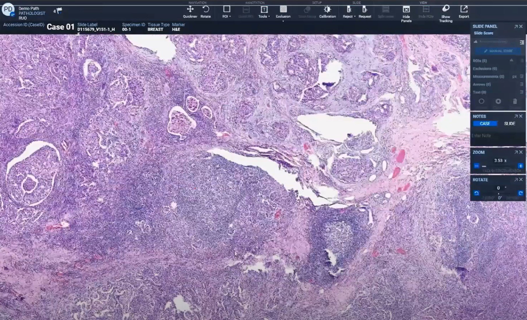
Of the many specialty areas for which clinical labs are implementing digital pathology, breast cancer is among the most common — and one of the applications where digital pathology has been the most helpful. The ability to perform whole-slide imaging has been valuable for samples with highly heterogeneous tissue and in light of the increasing complexity in the analysis of old and new biomarkers.
In a recent webinar, pathologists shared their experience with digital pathology tools for breast cancer analysis. If you’re considering this approach for your own lab–for breast cancer or any of the other areas where digital pathology is showing utility and benefits–the following highlights from the event may be useful.
More complex biomarkers, faster interpretations
For patients with breast cancer, new treatments have offered significant improvement in outcomes. But many of these therapies will only work for patients whose tumours have certain biomarker profiles. With each new therapy or combination of therapies, this puts pressure on clinical lab teams to perform deeper analyses and hence more time-consuming interpretation of each sample.
With more biomarkers to stain and track, digital pathology has been an important development, said Dr Richie Jara-Lazaro, Regional Pathologist in the Medical & Scientific Affairs division at Roche Diagnostics Asia Pacific.
Some digital pathology platforms offer analytical software that can also speed up the interpretation process. Computer-assisted scoring of gene copy numbers in a designated region of the slide image is faster than the standard manual process. Image analysis algorithms of the HER2 dual ISH assay, for example, can be used to determine HER2 gene amplification. According to Dr Jara-Lazaro, it might take 15 minutes to manually analyse a sample but only a few seconds with computer-assisted results.
At the Hospital del Mar in Spain, a pathology team led by Dr Mar Iglesias has implemented this approach. They began with breast cancer and found the visual heatmaps and computer-counted nuclei data to be so useful that they quickly expanded the use of digital pathology to other pathology subspecialties. Digital tools allow for precise tumour measurements and distances to margins that are not always possible with microscopy.
Streamlined tumour boards
According to Dr Iglesias, one of the reasons digital pathology has been so helpful is that it improves the results her team can share for tumour boards. For example, the digital approach allows them to annotate images and go back to them at any point, so they can be brought in as references at tumour boards as well as educational programmes whenever needed. For her clinician colleagues participating in these discussions, Dr Iglesias said, “digital makes their life easier.”
Pairing digital pathology results with a software-based tumour board interface also allows the clinical lab team and their hospital colleagues to assemble all clinical and pathology information into a single integrated system. This approach is more efficient and standardised, offering benefits for physicians and patients alike. Dr Iglesias said. “In fact, we are increasing the quality of this personalised care,” she added.
Digital pathology also improves collaborations and enables consultations because images can be shared easily within hospitals and across institutions, Dr Jara-Lazaro said. This enables remote or virtual tumour boards, which have been important for maintaining optimal patient care during the COVID-19 pandemic.
Implementing digital pathology ‘bit by bit’
When implementing digital pathology, one of the most important things to keep in mind is to stage the transition. “I recommend doing it bit by bit,” said Dr Iglesias, whose team first implemented digital pathology for breast cancer cases and has since rolled it out for pulmonary, skin and gastrointestinal cases, among others.
Adopting digital pathology requires considering the whole workflow: image acquisition, management, and image analysis. Ensuring that the lab has sufficient digital storage is as important as providing appropriate training to pathology staff who will have to shift from a manual to a digital technique.
At the Hospital Del Mar, one technician was designated to handle slide imaging when the new digital pathology workflow was brought in. That lab tech felt comfortable with the new process in a few weeks, said Dr Iglesias, pointing out that leading pathologist associations recommend a two-month learning period for people starting out with digital pathology tools.
From image analysis algorithms to software-enabled tumour boards, breast cancer provides some great opportunities for getting started with digital pathology tools. As more tests become available on digital pathology platforms, the dream of truly personalised medicine becomes increasingly within reach.


