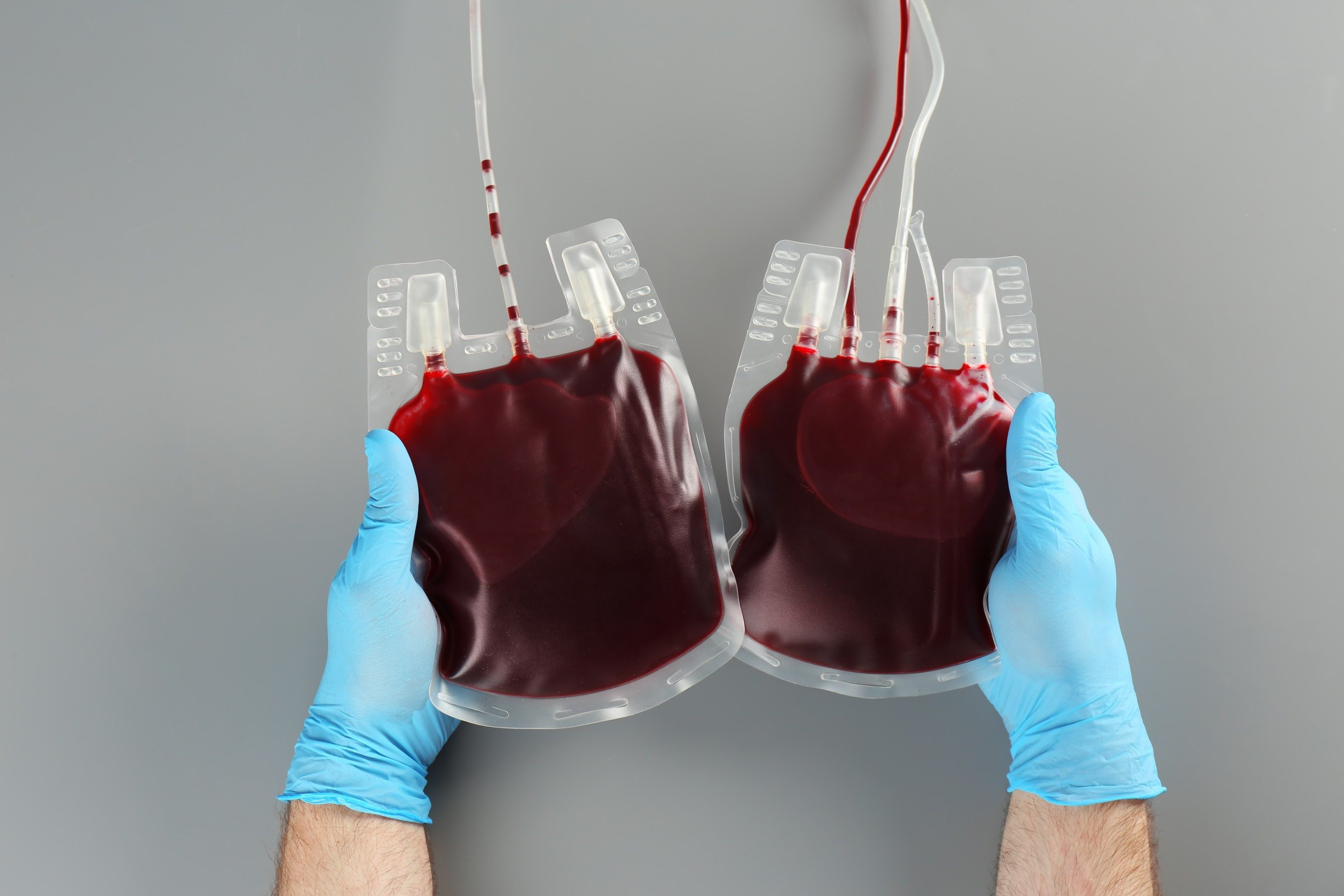
As part of their ongoing efforts to ensure a safer blood supply for patients, blood banks around the world are increasingly implementing screening systems for hepatitis E virus (HEV), a common infection that can lead to potentially serious and life-threatening outcomes in some patients.
In Thailand, the Thai Red Cross Society’s National Blood Center implemented a monthly HEV screening process at a rate of 500 units per month for the fiscal years 2023-2027. This initiative aims to mitigate the risk of HEV infection—particularly among pregnant patients, those with compromised immune systems, and other patient cohorts who are at greatest risk of poor outcomes from HEV infection.
In this report, we will look at the overview and epidemiology of HEV to understand why it is an issue that some blood banks have chosen to address, as well as how the screening process works, including considerations around laboratory technology and policy. We will conclude with some data from Thailand that explains the Thai Red Cross Society’s decision to implement HEV screening.
Overview and epidemiology of HEV
HEV is an RNA virus of the positive-sense, single-stranded, non-enveloped type in the genus Orthohepevirus, family Hepeviridae. The virus has at least four different genotypes: genotypes 1, 2, 3, and 4. Genotypes 1 and 2 infect humans, while genotypes 3 and 4 circulate in several animals, including pigs, wild boars, and deer. Occasionally, genotypes 3 and 4 infect humans [1].
According to data from the World Health Organization (WHO), approximately 20 million HEV infections have been newly diagnosed each year worldwide, leading to an estimated 3.3 million symptomatic cases [1]. The highest prevalence is in South and East Asia. In 2015, WHO estimated 44,000 deaths due to HEV infection [1].
These are four major routes of transmission of HEV infection: (1) fecal-oral transmission via the consumption of contaminated food and water; (2) the ingestion of undercooked infected animal meat; (3) vertical transmission; and (4) transfusion of infected blood products, a condition known as transfusion-transmitted HEV infection (TT-HEV infection) [1].
The incubation period of HEV infection ranges from 2 to 10 weeks, with an average duration of 5 to 6 weeks. Most patients with HEV infection are asymptomatic. Symptoms and signs of HEV infection can include fever, anorexia, nausea, vomiting, abdominal pain, itching, skin rash, arthralgia, jaundice, and hepatomegaly. These symptoms typically last 1 to 6 weeks. The immunity against HEV will be developed after the clearance of acute infection. In the majority of the patients, infection is self-limiting [1].
In rare cases, acute HEV infection can be severe and lead to fulminant hepatitis (acute liver failure). It can also cause high mortality and fetal loss in pregnant women with HEV infection, especially those in the second or third trimester. Moreover, HEV can cause chronic infection in immunosuppressed individuals such as patients living with human immunodeficiency virus (HIV) and organ transplant patients on immunosuppressive drugs [1].
HEV screening at blood banks: process and policy
Due to the fact that the majority of HEV-infected patients are asymptomatic and unrecognised, they might consequently donate blood. The prevalence of HEV infection among blood donors in previous studies ranges from 1:600 to 1:74,131 [2].
Several countries have implemented the universal or partial screening for HEV in blood donors, depending on the prevalence of HEV infection among blood donors within each specific area. Nucleic acid amplification testing (NAT) is the screening method of choice for HEV infection because infected blood donors frequently possess normal levels of liver enzymes and display negative results for anti-HEV IgM and IgG.
Most patients with TT-HEV infection are asymptomatic. Mild elevation of liver function tests might be present, leading to the delayed treatment. However, a few patients could develop signs and symptoms of hepatitis after receiving infected blood and blood products. Immunosuppressed patients are also more vulnerable to develop chronic HEV infection.
Currently, the European Association for the Study of the Liver Clinical Practice Guidelines on HEV infection recommend testing for HEV in patients with abnormal liver enzyme levels after blood transfusion [2]. Meanwhile, the routine screening for HEV in blood donors using the NAT method should be assessed based on the local risk of TT-HEV, along with a study of cost-effectiveness before any policy changes are implemented [2].
Global data and perspectives on TT-HEV
From a previous study in the South of England [3], 225,000 blood donors were screened for HEV RNA. Seventy-nine infected blood donors with genotype 3 HEV were identified with a prevalence of 1 in 2848 (0.04%). Among these infected donors, 62 blood products were transfused to 60 patients prior to the detection of the infection. Of 43 follow-up patients, 18 (42%) cases were infected. Transaminitis was common in TT-HEV infected patients. In order to successfully clear TT-HEV infection, two patients required treatment with ribavirin, and one patient had to reduce their immunosuppression. Furthermore, 10 patients had prolonged infection.
In Hokkaido, Northern Japan, there were 20 cases of patients with TT-HEV infection over a 17-year period [4]. Most of the infections were attributed to HEV genotype 3, with two patients infected by HEV genotype 4. Of 19 patients with available data, 9 had hematologic malignancies. The minimal infectious dose of HEV for TT-HEV infection in this study was 3.6 x 10⁴ IU HEV RNA. The estimated infectivity of HEV-contaminated blood products for transfusion was 50%.
Another study showed 0.011% (289 out of 2,638,685 blood donors) frequency of HEV infection in blood donors from 2005 to 2019 with the 20-pool NAT [5]. However, the frequency increased to 0.043% (597 out of 1,379,750 blood donors) when using individual NAT testing. Of the infected blood donors, 89% exhibited HEV genotype 3, while the remainder demonstrated HEV genotype 4. Since the implementation of HEV screening on blood donors, no further cases of TT-HEV infection have been reported (data up until 2019).
Updates from Thailand
In Thailand, the prevalence of HEV infection in blood donors was determined by HEV RNA at the National Blood Centre and Regional Blood Centre as shown in Table 1 [6].
Based on the data, the prevalence of HEV among Thai blood donors is relatively high, particularly within the regional blood service sector. This is why the National Blood Center has implemented a monthly screening process for Hepatitis E virus at a rate of 500 units per month (for the fiscal years 2023-2027).
References:
[1] https://www.who.int/news-room/fact-sheets/detail/hepatitis-e. Hepatitis E 20 July 2023.
[2] EASL Clinical Practice Guidelines on hepatitis E virus infection. Journal of hepatology. 2018;68(6):1256-71.
[3] Hewitt PE, Ijaz S, Brailsford SR, Brett R, Dicks S, Haywood B, et al. Hepatitis E virus in blood components: a prevalence and transmission study in southeast England. Lancet (London, England). 2014;384(9956):1766-73.
[4] Satake M, Matsubayashi K, Hoshi Y, Taira R, Furui Y, Kokudo N, et al. Unique clinical courses of transfusion-transmitted hepatitis E in patients with immunosuppression. Transfusion. 2017;57(2):280-8.
[5] Sakata H, Matsubayashi K, Iida J, Nakauchi K, Kishimoto S, Sato S, et al. Trends in hepatitis E virus infection: Analyses of the long-term screening of blood donors in Hokkaido, Japan, 2005-2019. Transfusion. 2021;61(12):3390-401.
[6] Pitchanan Khamsawang, Wilawan Sakram, Peeraya Suriya, Duangnapa Intarasongkrau, Patcharida Wongkittikul, Chompunekh Tungpunya, et al. Screening for Hepatitis E Virus Infection in Blood Donors. 2023.


